“`html
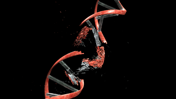
DNA injury detected in the brains of individuals afflicted with CTE might shed light on the disease’s presentation.(Image credit: circotasu/Getty Images)ShareShare by:
- Duplicate link
- X
Share this article 0Engage in the dialogueFollow usInclude us as a favored source on GoogleNewsletterRegister for our newsletter
The cerebral ailment chronic traumatic encephalopathy (CTE) has been associated with physical harm to the head — and it seems those cranial impacts may set off inflammation and DNA impairment that accumulates in cerebral cells over time, a recent study reveals.
The DNA deterioration, potentially culminating in cellular malfunction and demise, mirrors the damage observed in the brains of Alzheimer’s disease patients, as the study implies.
You may like
-

‘Chemo brain’ might result from injury to the brain’s elimination mechanism
-
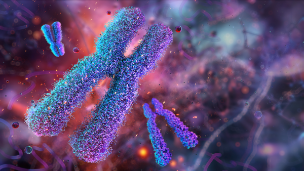
Gene located on the X chromosome could shed light on elevated multiple sclerosis prevalence in women
-

Insomnia and unease correlate with a depleted defense system — a recent study commences to disentangle the reasons
The scientific experts conducting the present study resolved to probe the association linking DNA deterioration and CTE after affirming the presence of accumulated mutations within mature neurons, which do not undergo division, throughout the lifespan. In a study from 2015, the team noted that these mutations intensify more rapidly amidst neurological disorders such as Alzheimer’s.
“We previously assumed that neurons maintained the most consistent genetic blueprints in the organism,” conveyed Dr. Christopher Walsh, a geneticist affiliated with Boston’s Children’s Hospital and a co-author for both the prior and present investigations. “However, it is evident that they accrue mutations annually, and the pace of these mutations accelerates in neurodegenerative diseases,” he elaborated to Live Science.
That discovery introduced an intriguing question: If DNA impairment escalates across various brain ailments, might it similarly precipitate neuronal attrition as witnessed in CTE?
In the new study, detailed in the Oct. 30 edition of the journal Science, scientists scrutinized the genomes of individual neurons obtained from 15 deceased individuals diagnosed with CTE, as well as those from four subjects with a past of frequent cranial impacts yet devoid of CTE. The group contrasted these neurons against cells derived from healthy brains and cells originating from individuals with Alzheimer’s disease. This process entailed single-cell whole-genome sequencing, a methodology that dissects all DNA material in each sampled cell.
The findings revealed that neurons extracted from CTE-afflicted brains exhibited a greater incidence of DNA mutations compared to neurons from healthy brains. On average, they presented roughly 114 supplementary single-letter variances in the DNA encoding per neuron. Conversely, neurons procured from persons exposed to repeated head trauma but lacking CTE did not manifest an increase in mutations when juxtaposed with healthy brains.
Researchers noted that the mutation pattern witnessed in CTE appears strikingly akin to the processes occurring in Alzheimer’s disease. Both conditions demonstrate a rise in the incidence of mutations alongside analogous categories of DNA shifts.
In the team’s former research, they “ascertained that neurons, despite not replicating, genuinely amass mutations at a continuous pace across their existence,” Walsh remarked. “Even within healthy brains, that progression advances by approximately 17 novel mutations each year from birth through advanced age. Nonetheless, within disease states, the progression intensifies.”
You may like
-

‘Chemo brain’ might result from injury to the brain’s elimination mechanism
-

Gene located on the X chromosome could shed light on elevated multiple sclerosis prevalence in women
-

Insomnia and unease correlate with a depleted defense system — a recent study commences to disentangle the reasons
The researchers also pinpointed an additional form of genetic impairment: abbreviated insertions and deletions, referred to as indels, where units are either appended to or eradicated from DNA’s framework. These microscopic DNA fractures were more bountiful in neurons from both CTE and Alzheimer’s brains contrasted with healthy ones. In select CTE instances, neurons held in excess of a thousand indels — equating to what might surface over more than a century of standard aging.
“These indels have escalated,” Walsh stated. “Their abundance likely suffices to instigate profound malfunction or demise within the compromised cells.”
Although the investigation abstained from explicitly assessing inflammation within the neurons, antecedent studies by study co-authors Dr. Ann McKee, a neuropathologist at Boston University (BU) CTE Center, and John Cherry, a neuroscientist at BU, have evidenced that widespread microglial activation — the brain’s immune operatives — in CTE brains typifies inflammation.
RELATED STORIES
—Laboratory-grown ‘minibrains’ aid in elucidating why traumatic cerebral harm elevates dementia susceptibility
—Whirling egg yolks imply how concussions distort the brain
—Years of persistent cranial impacts amplify CTE vulnerability — irrespective of being concussions
“We posit that CTE may represent a synthesis of recurring head trauma and inflammation,” Walsh articulated. “This amalgam may bombard the genome with analogous injurious processes as ultraviolet exposure effects on skin or tobacco consumption exerts on the lungs,” given both UV and tobacco engagement instigate DNA harm.
In summation, frequent cranial impacts may initiate inflammation within the brain, which can foster the accumulation of DNA mutations in neurons and facilitate cellular malfunction and demise. These conclusions imply that despite head trauma preserving its status as a critical precipitant of CTE, the enduring detriment is plausibly perpetuated by inflammation-induced DNA impairment.
The team currently explores whether analogous mechanisms unfold in additional neurodegenerative conditions, encompassing amyotrophic lateral sclerosis (ALS) and Huntington’s disease.
“This could signify a prevalent concluding trajectory across ailments,” Walsh suggested. “We aspire to map out the biochemical stages from inflammation to neuron expiration and discern potential intervention junctures.”

Larissa G. CapellaLive Science Contributor
Larissa G. Capella operates as a science writer anchored in Washington state. She secured a B.S. in physics along with a B.A. in English creative composition in 2024, thereby equipping her to pursue a vocation integrating both disciplines. She predominantly provides coverage on environmental, Earth-based, and physical sciences, though she remains receptive to authoring pieces pertaining to any scientific domain sparking her inquisitiveness. Her work has manifested in Eos, Science News, Space.com, among other outlets.
Show More Comments
You must confirm your public display name before commenting
Please logout and then login again, you will then be prompted to enter your display name.
LogoutRead more

‘Chemo brain’ might result from injury to the brain’s elimination mechanism

Gene located on the X chromosome could shed light on elevated multiple sclerosis prevalence in women

Insomnia and unease correlate with a depleted defense system — a recent study commences to disentangle the reasons

Heart attacks are less harmful at night. And that might be key to treating them.

Aging and inflammation may not go hand in hand, study suggests

We may finally understand stress-induced hair loss
Latest in Neuroscience
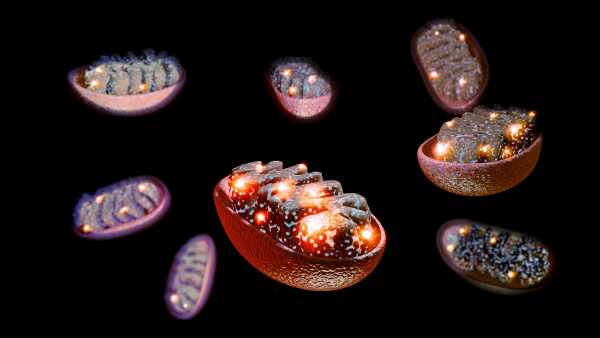
‘Mitochondrial transfer’ into nerves could relieve chronic pain, early study hints