A 28-year-old sorceress professes to have unraveled the infamous 1945 Charles Walton homicide in Lower Quinton, Warwickshire, by communicating with the deceased’s spirit. Rosie asserts the vicious slaying concerned finances and coercion, not necromancy. Share Article Share Article Facebook X LinkedIn Reddit Bluesky Email Copy Link Link copied Bookmark Comments

A decades-spanning killing persists in casting an ominous presence over a modest Warwickshire town, with law enforcement, detectives and amateur sleuths still unable to ascertain who heinously murdered a cherished farmer.
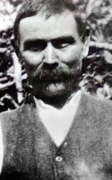
Now, Rosie, a 28-year-old enchantress, professes to possess the solutions – and her inferences are unsettling. The psychic purported she summoned Charles Walton, 74, the casualty of the barbaric ‘Witchcraft killing’ assassination in 1945. The homicide was appalling.
Article continues below ADVERTISEMENT
Charles’ digging implement was thrust through his artery and his physique was secured to the soil with his hay fork. He also bore a sizable cross etched into his delicate chest.
The cross mirrored slayings linked to devilry – igniting conjecture over the participation of magic, states the Daily Star.
Nevertheless, Rosie is persuaded no devilry was implicated in the assassination and this was utilized as an “evasion maneuver.”
Article continues below ADVERTISEMENT
READ MORE: ‘I’m a private investigator — these are the places I check for cameras’
READ MORE: Woman gang-raped in popular holiday spot as 3 Brits face decade in jail

Alternatively, she stated a “masculine vitality intent on harming” Charles, a Lower Quinton farmhand who occupied a leased residence across from the village church alongside his niece, Edie.
Article continues below ADVERTISEMENT
Charles labored on regional farms until the day of his demise. He was well-liked although recognized for being peculiar.
He would routinely transport feed and untamed avians would assemble on his hand, feral canines would also be pacified merely by his tone.
On this account it was alleged he was intertwined with sorcery or dark wizardry. Tragically, Charles was killed on Valentine’s Day as he was maintaining hedges on Hillground.
Amid the inquest, an officer happened upon a tome he regarded as potentially germane. The volume, Folklore, Old Customs and Superstitions in Shakespeareland, inscribed by J.
Harvey Bloom in 1929, encompassed a chilling excerpt detailing the demise of another Charles Walton who perished in 1885 – 60 years prior to this Charles – subsequent to a spectral encounter.
The matter amassed such notice that Scotland Yard’s Detective Inspector Robert Fabian was summoned, yet even he proved unable to decipher the enigma.
Rosie probed into the ominous influences at play in this disquieting assassination and how she arrived at her individual deductions.
She recounted: “I sat with this last week and permitted everything to materialize and let spirit permeate because it was very intricate. I can appreciate why this was never explicitly resolved because it was a profoundly complex scenario.”
Indeed, the hamlet endures as taciturn regarding the assassination to this present day, with inhabitants declining to deliberate it.
Rosie declared: “I wish to commence by conveying necromancy was employed as an evasion stratagem – and the genuine assassination didn’t transpire owing to any necromantic participation, it genuinely transpired nearer to domestic affairs and it transpired in relation to finances.
“There was a monetary predicament that unearthed some virulent vitality and I perceive the victim was being extorted somehow and they attained a juncture where they could no longer satiate that anymore and that’s the justification they were assassinated.
Don’t miss: Chilling video shows squalid ‘prison cell’ where ‘witch’ kept woman as slave
Don’t miss: ‘Diabolically chilling’ horror movie ‘is perfect in every way’ on Film 4 tonight
“I genuinely surmise this is a male who assassinated him and I genuinely surmise it was someone in close proximity who was harboring something against Charles.
“There was considerable concealment surrounding Charles – he mingled in a manner that he didn’t reveal overly much and I perceive he possessed veiled aspects to him.
“Even when I was communicating with the culprit I sensed they were vastly misunderstood and because they were, their dispositions were exploited and there were specific things this individual perpetrated which intimidated other individuals which is why they wielded finances over Charles’ head.
“I’d be intrigued to ascertain if there was something concerning land and territorial inconsistencies that had greater involvement to familial affiliations. I genuinely surmise that there were also domestic quandaries that stretched back that were unearthed further down the track.”
The latest science news from the natural world, health, tech and beyond Subscribe Invalid email
We use your sign-up to provide content in ways you’ve consented to and to improve our understanding of you. This may include adverts from us and 3rd parties based on our understanding. You can unsubscribe at any time. Read our Privacy Policy

Get More of Our News on Google
Set Daily Express as a ‘Preferred Source’ to get quicker access to the news you value.

The proprietor of the village tavern, the College Arms, Tony Smith, communicated to the BBC he could articulate nothing. He stated: “I can’t converse with you regarding that. After 17 years of supervising this establishment, I am aware there are certain matters we don’t deliberate.”
The property altercations and sinister masculine presences Rosie delineates could readily be the undercurrent maintaining Lower Quinton hushed – but until somebody breaches their quietude – we may never unearth how Rosie’s revelations align with the actuality.