Using modern technology, scientists have “digitally unpacked” 2,000-year-old animal mummies from Ancient Egypt and discovered the causes of their death.

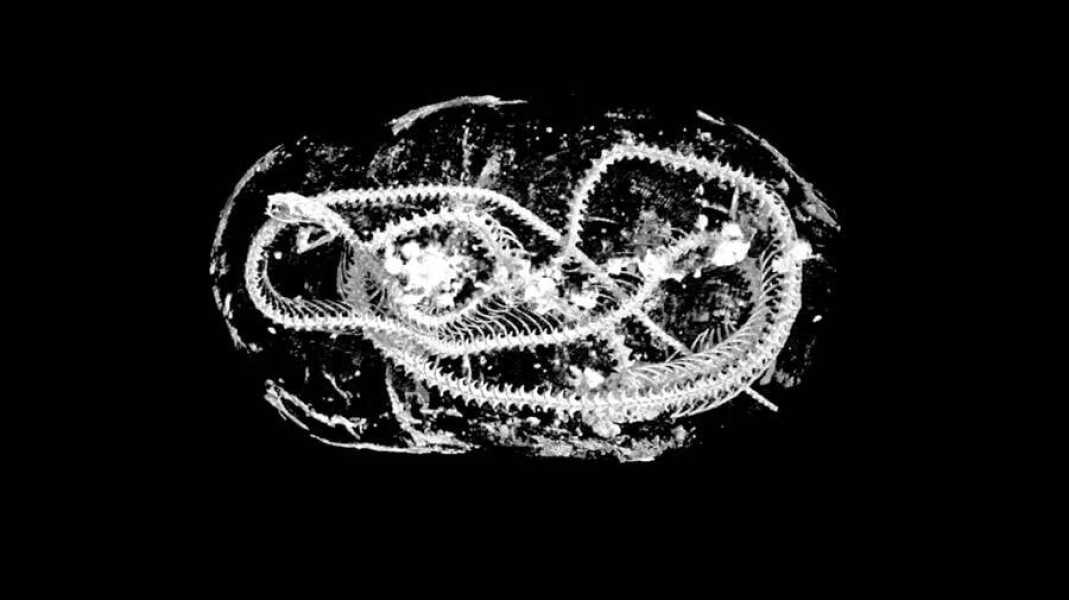
Swansea University Micro-CT scan of a mummified Egyptian cobra dating back to 2000.
A team of researchers from Swansea University in Wales have studied the remains of mummified animals from Ancient Egypt. The study was carried out without damaging the artefacts using modern high-resolution 3D digital scanners, which led to a “digital unpacking” of the ancient remains.
According to Gizmodo , the mummified animals date back 2,000 years. A study of their embalmed remains has provided scientists with a wealth of information about how these creatures could have lived and died so long ago. The study's findings were published in Scientific Reports .
The subjects of the study were the wrapped bodies of a bird, a cat and a snake. Although using X-ray scanners to study ancient mummies has become a fairly common practice among archaeologists, the team of researchers in this case used micro-computed tomography, also known as micro-CT scanning.
The advantage of this technology is that it provides exceptionally high-quality images that are 100 times more detailed than images from a conventional medical CT scan. Moreover, these images also offer 3D measurements of objects. The technology is so precise that it allowed the team to examine the teeth of mummified animals.
“Using micro-CT we can effectively perform autopsies on these animals more than 2,000 years after they died in ancient Egypt,” said Richard Johnston, a professor at Swansea University and leader of the study.
“These are cutting-edge scientific imaging techniques. Our work demonstrates how today’s high-tech tools can shed new light on the distant past.”
Johnston and his team were able to extract significant information about the wrapped animals thanks to the exceptionally detailed images produced by micro-CT.
“I chose a few samples with different shapes to demonstrate this technology, not knowing what we would find at that stage,” Johnston told Gizmodo about his approach to choosing samples for the study.
“So the mummies of a cat, a bird and a snake were chosen. There are many examples of these mummified animals in museums and they have been studied throughout history. We wanted to test the limits of what this technology could reveal that had not been possible before.”
Micro-CT scans of the kitten's mummy revealed that it was a pet that died when it was less than five months old, which the researchers were able to determine by “slicing” virtual scans of the kitten's jaw. This information had not been noticed by previous researchers when analyzing 3D data on a 2D screen and in a 3D print.
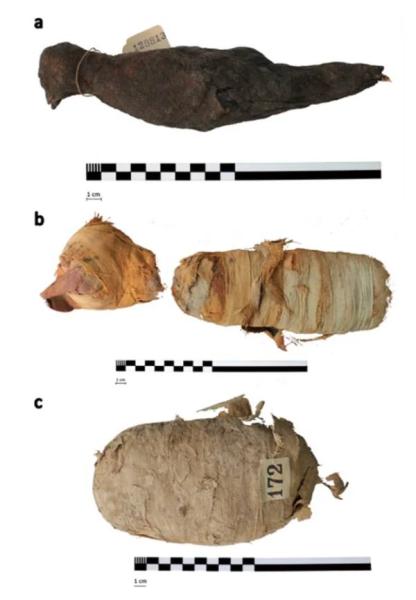
Swansea University. Appearance of mummified animals: birds (a), cats (b) and snakes (c).
The kitten's neck was also broken, either shortly before its death or just before mummification, to hold its head upright for embalming.
As for the snake mummy, the team found that it was a young Egyptian cobra that had developed a form of gout, likely as a result of dehydration during its life. The spinal fractures
Sourse: www.allthatsinteresting.com