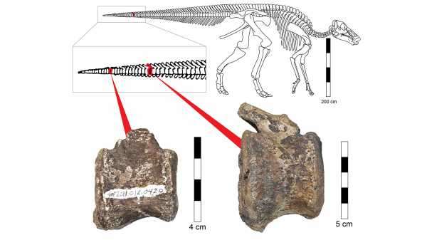

Two tailbones in a juvenile hadrosaur — a duck-billed dinosaur — bear telltale cavities that hint at tumorous growths.
A rare disease that causes tumors in humans produced similar growths in dinosaurs, new research suggests.
Scientists discovered unusual circular cavities in a pair of tailbones that belonged to a hadrosaur, or duck-billed dinosaur, found in Dinosaur Provincial Park in Alberta, Canada. In shape and structure, the lesions closely resemble scars created in human bones by growths caused by Langerhans cell histiocytosis (LCH), a disorder in which excess immune system cells build up as benign tumors, according to the new study.
(NLM). These tumors mostly affect children between the ages of 2 and 10, and though the growths are benign, they can cause swelling and pain; in severe cases they may fracture the bone from the inside.
Medical researchers have studied LCH since the 19th century. However, some controversy remains about whether LCH is technically a cancer, as the tumors consist of only a few abnormal cells surrounded by lots of normal immune cells, representatives of the Histiocyte Society Scientific Committee said in a statement.
Cavities that LCH tumors carve into bone are “well-defined” pits with a distinctive shape — columns along the walls and wrinkles at the base, according to the study. The lesions in the hadrosaur bones “were different in appearance from those seen in metastatic cancer, tuberculosis, and fungal disease,” and were most similar to LCH lesions in human bones, the scientists wrote in the study.
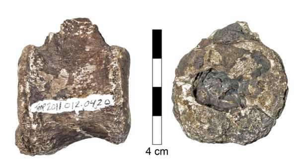
Circular depressions in the hadrosaur vertebrae were “very similar” to lesions caused by LCH in people.
A unique shape
The study authors examined the lesions in the hadrosaur bones using microscopy and computerized tomography — CT scans — which combines multiple X-rays to create a 3D digital reconstruction of an object. This allowed the researchers to visualize tiny blood vessels that fed the tumors, and thereby reverse engineer the long-vanished growths, said study co-author Hila May, a lecturer with the Department of Anatomy and Anthropology in the Sackler Faculty of Medicine, at Tel Aviv University in Israel. May led the team that scanned the hadrosaur bones and reconstructed the tumors.
When viewed in macro resolution, the holes expanded into the surface of the dinosaur vertebrae in a shape that was “very unique. We don’t see it in other tumors that we know from humans,” May told Live Science. “This opening toward the surface is very typical to LCH, and that was the first clue.”
Another important piece of evidence was the damage to the bone’s microstructure, which formed a pattern that is also commonly seen in LCH tumors’ cavities, May said. The researchers also analyzed human bones: some with LCH lesions and others with cavities and scars caused by other pathologies. When they compared those tumor scars with the hadrosaur lesions, the closest match was the cavities caused by LCH, the study authors reported.
“After we saw that, we could give the most probable diagnoses of this lesion — which is LCH,” May told Live Science. “And it’s very similar to LCH in humans.”
Identifying and studying diseases that affect people and non-human animals alike could help scientists to better understand the environmental factors that shape these illnesses, “which maybe, in the future, will give us a clue about the cause — or the solution,” May said.
The findings were published online Feb. 10 in the journal Scientific Reports.
Sourse: www.livescience.com