Paleontologists have described an infectious lesion of the air sacs in a young sauropod that lived about 150 million years ago. The disease was most likely caused by bacteria or fungi. As noted in an article for the journal Scientific Reports, air sac infections often affect modern birds, but non-bird dinosaurs have not yet been so diagnosed.
Evidence of infections and other diseases is poorly preserved in the fossil record. Nevertheless, paleontologists sometimes manage to diagnose representatives of species that went extinct millions of years ago. For example, specialists recently for the first time identified cancer in a non-bird dinosaur and, a few years before that, infectious arthritis. In addition, there are numerous examples of parasitic infections from which extinct animals suffered. You can read more about them in our article “Passenger from the Mesozoic”.
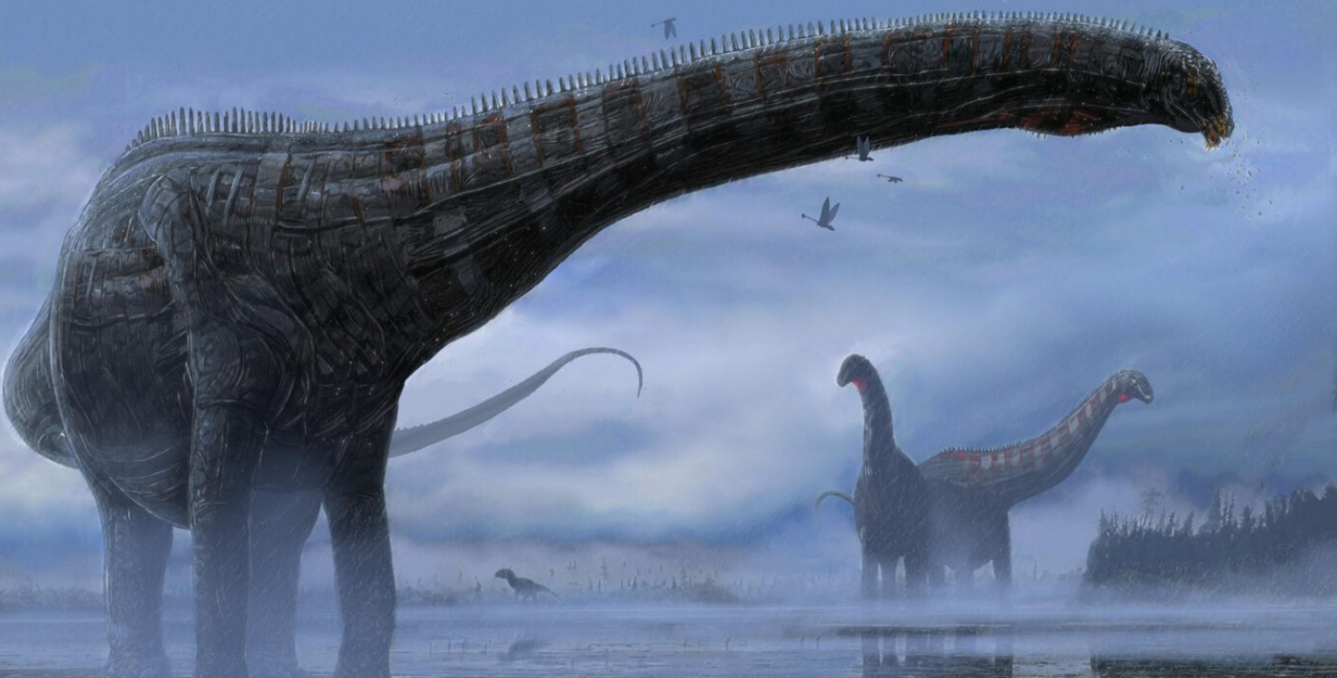
A team of paleontologists led by D. Cary Woodruff of the Great Plains Dinosaur Museum has demonstrated that non-bird dinosaurs faced another group of diseases. The researchers focused on the remains of an immature sauropod from the Diplodocinae clade, 156.3-146.8 million years old, found on the Late Jurassic Morrison Formation in Montana. The specimen, tentatively named MOR 7029, was collected in several stages: a skull and seven articulated cervical vertebrae were discovered in 1990, and additional postcranial skeletal elements were found in 2013-2015.
On close inspection of the remains of MOR 7029, Woodruff and colleagues noticed evidence of an unusual pathology. On the surface of the air cavities (the sauropods had air sacs inside them, similar to those found in modern birds) of the fifth to seventh cervical vertebrae, the bone tissue strongly overgrew, forming a wavy and heterogeneous texture. The largest focus of overgrowth arose in the left ventral cavity of the fifth cervical vertebra. Its dimensions were 2.8 centimeters in the head-to-tail direction and 2.1 centimeters in the dorsoventral direction. At the same time, the height of the outgrowths deep into the cavities varies from half a centimeter to a centimeter.
Using computed tomography, the researchers also identified numerous defects within the affected vertebrae. These are mainly small cavities of ovoid, tubular and angular shape. They are unevenly distributed throughout the vertebrae and are more concentrated around foci of bone overgrowth. These signs indicate serious deviations from the normal pneumatization of the vertebrae. In modern species, similar problems occur with chronic disease.
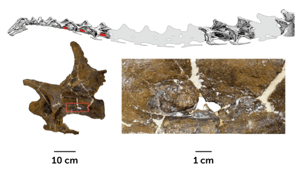
A. The skeleton of the head and neck MOR 7029. Affected vertebrae are marked in red. B. Fifth cervical vertebra MOR 7029, affected area highlighted in red rectangle. C and D. The affected area in close-up.
Paleontologists often find sauropod vertebrae with various pathologies. Until now, however, no specialists have described the lesions found in MOR 7029. Because the foci of bone overgrowth formed on the surface of the air cavities, Woodruff and colleagues suggested that the young sauropod’s ailment was localized in the air sacs. To understand what kind of disease MOR 7029 suffered from, the authors made an analogy with birds – the only surviving group of dinosaurs.
The researchers considered the cancer version unlikely. The fact is that malignant tumors rarely develop in the air sacs of modern birds. The clinical picture of the disease MOR 7029 also differs from that typical of cancer. According to an alternative hypothesis, the air sacs of sauropods became inflamed due to inhalation of dust, for example, during a volcanic eruption. This condition is called pneumoconiosis. However, some of the dinosaur’s symptoms are not consistent with this diagnosis either.
In the end, Woodruff et al. settled on the idea that MOR 7029 likely suffered from aerosacculitis – an infectious lesion of the air sacs, the researchers admit that making an accurate diagnosis for an animal that died about 150 million years ago, it is almost impossible. Nevertheless, the infection hypothesis most economically explains all the available facts. In that case, this is the first known case of aerosacculitis in a non-bird dinosaur.
The causative agent of the disease identified in MOR 7029 could be mycobacteria, chlamydia, or fungi of the genus Aspergillus. All of them still frequently cause aerosacculitis in birds today. If respiratory infections manifested themselves in dinosaurs in the same way as in modern feathered birds, the young sauropod suffered from cough, runny nose, fever, lethargy, diarrhea, and weight loss. It is possible that these symptoms are what killed him.