
The new embryo atlas was developed using serial images of zebrafish embryos developing under a microscope. (Image credit: Royer Group, CZ Biohub San Francisco)
Amazing new psychedelic videos provide a glimpse into what living organisms look like in the earliest moments of their existence – something that took scientists years to discover.
The videos are part of a new embryo atlas called Zebrahub, which shows where cells are located and what they do at different stages of development. The atlas combines high-quality time-lapse videos of developing embryos with data showing which genes are active at each stage.
The atlas covers embryos of zebrafish (Danio rerio), a fish often used in biological research. Most of the genes of these small fish have close analogues in humans, and the main components of the cells are common to the entire vertebrate branch of the tree of life.
Zebrahub: New perspectives on developmental biology – YouTube

See more
“At these early stages of life, all embryos are very similar,” said Loic Royer, one of Zebrahub’s developers, head of the Organismal Architecture group and director of AI visualization at Chan Zuckerberg Biohub San Francisco. “The shapes, the genes, the molecular machinery that goes into making the organism — it’s all very similar.”
Royer is the senior author of a new paper describing Zebrahub, published Thursday (October 24) in the journal Cell. He added that it’s hard to predict what discoveries the new tool might yield, but studying embryos from other species could help answer questions about how birth defects and other disorders arise in humans. What’s more, the new atlas could provide clues about why animals like zebrafish are able to regenerate body parts after injury while we can’t, he speculated. It could also reveal significant differences between young and aging tissues, which could help explain why aging occurs.
At its core, Zebrahub is focused on one central question. “It’s fundamentally about how we’re wired,” Royer told Live Science. “If we don’t understand how we’re wired, how can we hope to ‘fix’ ourselves?”
Zebrahub is freely available and offers tools that allow biologists to explore and analyze vast amounts of data. However, to collect the initial data, Royer and his team had to develop new methods for studying zebrafish embryos.
Historically, research has focused on either where cells are located in a developing embryo or which genes are currently being activated. To track where cells are, scientists take multiple pictures of the embryos under a microscope. Zebrahub's developers have created a new microscope that runs a thin line of light across the embryo, creating images as it moves. This technique avoids exposing the embryos to harsh laser light, which can damage them.
Image 1 of 4
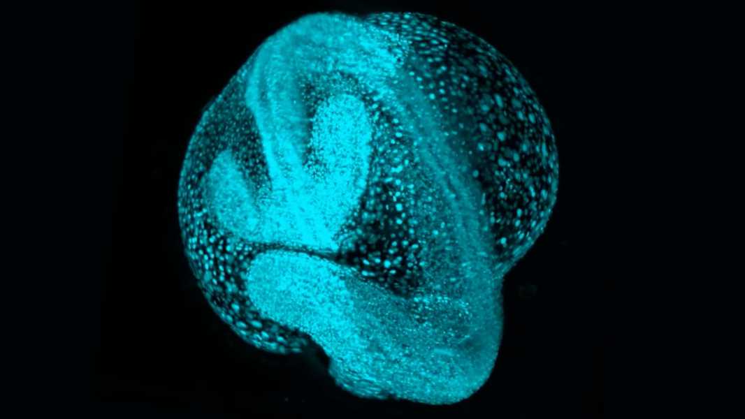
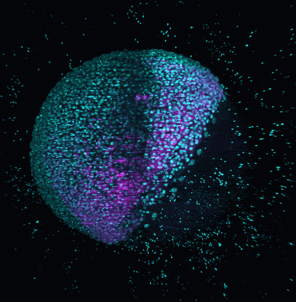
This image shows cells from an early zebrafish embryo. (Photo courtesy of Royer Group, CZ Biohub San Francisco)
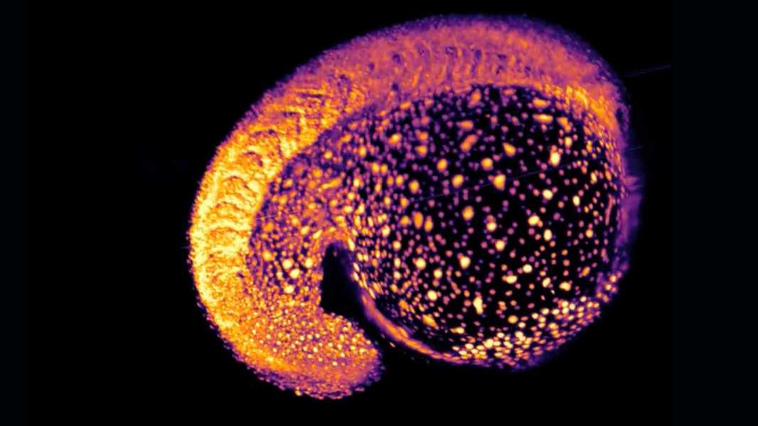
This image shows cells from an early zebrafish embryo. (Photo courtesy of Royer Group, CZ Biohub San Francisco)
Sourse: www.livescience.com