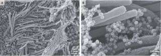

Images of SARS-CoV-2 captured with a scanning electron microscope. The SARS-CoV-2 virus particles appear as tiny, ball-looking structures. The noodle-like projections in the images are cilia, or hair-like structures on the surface of some airway cells.
Striking new images show a close-up view of the novel coronavirus as it invades cells that line the human airway. The images provide a glimpse of the staggering number of virus particles that are produced during an infection — indeed, infected cells can churn out virus particles by the thousands.
Camille Ehre, an assistant professor at University of North Carolina School of Medicine’s Marsico Lung Institute, captured the images with a scanning electron microscope, a device that uses a focused beam of electrons to produce images. To obtain the images, published Wednesday (Sept. 2) in The New England Journal of Medicine, researchers first infected human airway cells with SARS-CoV-2 — the coronavirus that causes COVID-19 — in lab dishes, and then examined the cells after four days.
The noodle-like projections in the images are cilia, or hair-like structures on the surface of some airway cells. The tips of the cilia are attached to strands of net-like mucus, which is naturally present in the airway.
Related
—20 of the worst epidemics and pandemics in history
—The 12 deadliest viruses on Earth
—11 (sometimes) deadly diseases that hopped across species
The SARS-CoV-2 virus particles look like teensy balls. At a high magnification, you can make out the spiky structures on the surfaces of the virus particles. Those crown-like spikes give the family of “coronaviruses” its name. (“Corona” means “crown” in Latin.) The virus uses those spikes to invade human cells.
The image also shows a high density of virus particles, or virions. An analysis of the concentration of virus in the sample suggests a “high number of virions produced and released per [airway] cell,” Ehre wrote.
Originally published on Live Science.
Sourse: www.livescience.com